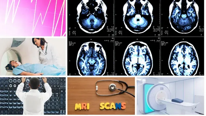
Understanding MRI Scan?
MRI stands for Magnetic Resonance Imaging. It's a diagnostic procedure that provides detailed pictures of the inside of the body using a powerful magnet, radio waves, and a computer.
An MRI uses very strong magnetic fields to align all nuclear spins. It then sends an RF pulse that flips tissue's natural polarization bias in different axes which refocuses protons magnetically along pre-determined axes of alignment. By flipping their inherent spin orientation during the readout process, scientists are able to create images presenting how molecules are oriented within living tissue or at specific points in time after an event has occurred. This technique is known as magnetic resonance imaging (MRI) because it causes nuclei atoms to respond with varying intensity to externally applied electromagnetic fields following established.
MRI or Magnetic Resonance Imaging is a radiology procedure that uses powerful magnets to create various images of the inside area of the body. It is important to know that MRIs are preferred in people with metal implants. We use MRIs to help diagnose things like cancer, spinal stenosis, joint pain, and more! We can also use MRI for many different types of chiropractic treatments such as pregnancy-related treatments, lower back pain relief therapy services. The MRI scanner or Magnetic Resonance Imaging device is a large and complicated medical machine that will allow the doctor to perform an in-depth scan of our internal organs.
An MRI scans your body by generating electromagnetic signals from a powerful magnet. Then it uses them to look for differences in the patterns of the energy emitted by different types of tissue. An image is produced using a computer program, which can highlight certain types of tissue because they respond differently to the scanned magnets' magnetic fields. This includes muscles, cartilage, fat, and bones - but not fluids or other tissues such as nerves or bowel contents. An MRI scan generates a three-dimensional image of the inside of the body. The patient lies on a parenthesis-shaped, hollow steel or lead magnet called an electromagnet while scanning coils are arranged around them. When the person is in the middle of it all, computer-controlled radio waves are used to create images.
Magnetic resonance imaging (MRI) is a medical imaging technique that uses strong magnetic fields and radio waves to produce high-quality two-dimensional diagnostic images of internal organs and soft tissues within living subjects noninvasively. ?1;
An MRI scan is a sequence of images that are taken from different angles around the body to produce a detailed internal picture or to look for abnormalities. MRI machines, often called "big magnets" because they create strong magnetic fields might also be used in imaging situations where X-rays can't provide sufficient detail, such as in the case of pregnant women and certain types of cancer patients.
An MRI (magnetic resonance imaging) scan uses a combination of radio waves and magnetic fields to take pictures through the body. MR scans use magnetism, radio waves and strong electromagnetic fields together to produce good quality images of the structures in your body that may or may not be visible externally. MRI scanners create detailed two-dimensional electronic images in slices from many different angles: like cutting a loaf of bread into thin, cross-sectional slices for examination. The machine creates images by using powerful magnetic fields which align the spinning nuclei atoms inside cells with one another temporarily into ferromagnetic alignment with its own axis perpendicularly - this then enables researchers to "see" what these nuclei are doing without needing contrast agents.
Essentially an MRI Scan is a medical imaging technique that uses the magnetic properties of radio waves to generate images of body structures. Often, MRI investigations are used to diagnose diseases including cancers, vascular lesions, bone abnormalities, and spinal disorders. It can also have applications in other areas such as prenatal care for pregnant women. MRIs use no ionizing radiation, but physicians should be aware that there are some risks associated with exposure to very large electromagnetic or radiofrequency fields, which include heating of tissue and induction currents in metal implants.
MRI scan is the layman's word for Magnetic Resonance Imaging scan. It allows us to see or measure different types of tissues in the human body like bone, tendon, cartilage, fat and other soft tissues (muscle). There are different parts of an MRI scan that assess different parts of the body; these include axial images (bone) coronal images (middle section) sagittal images (top view), 3D images, and more complex scans like cine sequence or gradient echo imaging to study joints. We choose what type of image we want by changing settings on the machine.
Magnetic resonance imaging (MRI) is a medical imaging technique used to obtain images of the body's internal organs. It does this by using powerful magnets and pulsed electromagnetic fields, which interact with atomic nuclei in your body. For an MRI exam, you lie on a table that slides into a long narrow tunnel-like scanning magnet that surrounds most of your body. This procedure is painless and lasts less than 30 minutes...
MRI stands for Magnetic Resonance Imaging. MRI scanners are large, doughnut-shaped magnets with computers in the middle that allow us to take pictures of things inside the body without cutting them open. MRI is very good at looking at brain disorders, torn ligaments and can even see bones that are under your muscles. It's also great for heart problems etc.
Miniature radio transmitters positioned just outside it generates radio waves that penetrate your body tissues creating an image of their structure within seconds. Then a computer digitizes all this information so it can be viewed on a monitor or printed out on film. “No harmful radiation is being used"* Magnetic Resonance Imaging (MRI) is done when non-invasive imaging, such as X-rays or ultrasound, are not helpful in showing the organs inside your body.
An MRI scan may be used to diagnose degenerative diseases like Alzheimer's disease and Parkinson's disease. The ability to produce highly detailed images of brain structures has also made it one of the most researched medical imaging techniques. Frequently there is a need for MRI scanning when relocating bone joints, assessing developmental abnormalities in children, determining brain injury after an accident or stroke, diagnosing herniated discs and tumors, cancer screenings. MRIs use magnetic fields and radio waves to learn about the inside of the body without surgery. Sometimes MRIs are used as part of a diagnostic toolbox that also includes X-rays, computed tomography scans (CTs), blood tests, and physical examinations. The results from those other evaluations can help determine if an MRI would be helpful for a diagnosis or treatment decision. For example, CT uses a different type of radiation to create images than does MRI scanning. Brain CT helps doctors assess things like bleeding inside the skull following
There's no requirement for an MRI scan. You can get one if you believe it will help your diagnosis. Your doctor would decide which radiologic study might be appropriate for you. Some conditions - like cancerous tumors - are understandable where imaging is considered to possibly provide information that guides management decisions, or simply to exclude possible causes of symptoms (since many benign causes involve changes in the same region). Additionally, contrast-enhanced CT scans and MRIs are helpful in making specific diagnoses once lesions have been found on physical examination; for example, they're often used to diagnose inflammatory bowel disease (IBD) including Crohn's disease and ulcerative colitis.
MRI scan is most often done to assess the causes of major abdominal pains, bowel blockages, headaches, bleeding in the brain, problems with joints and muscles, tumors of unusual size on an X-ray or CT scan. MRI Scans are also very helpful for looking into problems that involve the abdomen or pelvic region. Conditions that produce changes in bowel habits or appetite can be readily diagnosed with an MRI. There are some cancerous tumors of certain types that are more easily seen with an MRI than any other type of imaging method because they do not show up well on x-rays or computed tomography (CT) scans.
MRI scans are usually required to tackle the following issues:
1) Hodgkin's Lymphoma
2) Brain Tumors
3) Cervical Spinal Cord Diseases
4) Frq-siissiiancy of Nerves, Muscles Or Joints
5) Sometimes Chest Disease, Liver Disease etc.
6) Carcinoma - To Find Out Whether The Cancer Has Spread.
An MRI Scan is used to look for abnormalities with structures inside the head, which can diagnose many different conditions. It may be used to detect tumors; blood clots that cause strokes; swollen or damaged brain tissue; infection of the covering around the brain (meningitis); wear and tear of the cartilage in your joints, like osteoporosis; deterioration of bones (osteoarthritis) The most common application is medical imaging of brains. MRI gives more detailed, higher resolution images than CT scanning because MRI employs both strong magnetic fields and radio waves. Images are produced by combining radiofrequency pulses with magnetism to produce signals on long hoses routed through an entire body.
MRI is examining what's going on inside your body by putting you in a big magnet, then only use the MRI scan to show us. During an MRI scan, you are placed within a large scanner called "the bore" which uses magnets and radio waves to line up the atomic particles in the body so they can be analyzed. Your doctor will recommend an MRI when there's something abnormal or unclear about your symptoms or an injury. An MRI talks more about soft tissues than X-rays do because X-rays pass through the skin easily but don't penetrate beyond muscle tissue; MRIs go deep into all organs of the body including breast tissue.
Catch it early while the tumor is still small and end up with a much better prognosis. As tumors grow, they disrupt normal tissue and cause cells to release chemical signals that attract blood vessels (angiogenesis or neovascularization) which can make it difficult to capture clear images. The American Cancer Society estimates that about 2,350 patients in America will die from lung cancer this year. The best way to save your life is by catching it early while the tumor is still small and end up with a much better prognosis. So, if you were just told you have late-stage lung cancer, understand that there's no chance for success without chemotherapy/radiation therapy combined because of the already advanced size of your tumor.
MRIs are used to diagnose abnormalities in fetal brain development, to study how trauma affects the brains of individuals with suspected head injuries, and for locating tumors or other health problems that might not show up on an X-ray or CT scan. MRI is often done before any surgery involving the brain. It's also recommended after a concussion. MRIs have come in very handy when it comes to diagnosing patients who have had strokes." They are also occasionally used in the emergency room setting when doctors need help placing an intravenous line where they can't find one by palpation alone." Conditions diagnosed with MRI include many types of pain in different parts of the body, blindness/visual disturbances, lesions found during workups for other conditions.
MRIs are not recommended for patients who have Eastern equine encephalitis (EEE), cerebral malaria, or viral encephalitis because the magnetic field can make them worse. Patients should also decline MRIs if they've had cancer within six months, MRI contrast dye exposure within six weeks, an implanted defibrillator, or pacemaker.