Diagnostics : Understanding PET Scan – When doctors recommend it & what are the disadvantages if any.
Why would a doctor recommend a PET scan
A PET scan is an imaging study that produces clearer, more detailed information about the body, especially when looking for disease. The most common PET scans are used to diagnose cancers and show tissue changes in different parts of the body such as lungs or kidneys.
A PET scan is often done if a patient is having symptoms like confusion, sudden weight loss, and vomiting. These symptoms can be signs of cancer but they can also be caused by other diseases like ulcers/peptic ulcer disease (indigestion), low blood sugar (diabetes), and chronic kidney or liver failure. A doctor will recommend a PET scan if these symptoms last longer than three months in order to find out what may be causing them.
The PET scan is recommended for many cancers to determine the viability of chemotherapy treatments, which can be too harsh for people with low levels of glucose in their cells.
Positron-emitting radioactive tracers like fluorodeoxyglucose (FDG) are used to label and identify tissues in organs in your body. The concentration FDG labeled products in a particular organ will show how active that organ is functioning in the body. For example, when cancer cells are active there is often an increased uptake FDG inside these cells (a person's cancerous tumor). With this information, doctors may recommend lower doses of chemotherapy which can be more tolerable for patients who have low blood glucose levels.
A PET scan can help a physician diagnose a variety of conditions, including lung/liver disease, cancer problems, coronary artery disease and stroke. It's also used to assess radiation exposure or detect physical injuries.
Modern nuclear medicine techniques use short-lived radioactive molecules called radioisotopes that are injected into the body to provide information about function in specific organs or tissues. The radioisotope is usually combined with an accompanying medication that is being tested for the problem being studied. In some cases, a not-radioactive compound can be inserted into a vein to produce an image like what will be seen in a PET scan in another laboratory without exposing patients to any radiation at all.
A doctor typically recommends a PET scan for any change in an individual's physical or mental health. A PET scan helps doctors diagnose cancers, neurological diseases, heart disease, lung disease, liver disease, and more. The test can be done at the same time as other tests that are already planned to be taken place, or it can be done on its own without warning.
A PET scan is recommended when the diagnosis of carcinoma, sarcoma or leukemia is uncertain, given atypical findings on any imaging exam done on the individual's digestive organs. PET scans are usually used on individuals with unexplained gastrointestinal issues.
Positron emission tomography (PET), sometimes called single-photon emission computed tomography (SPECT) involves using gamma rays that pass through tissue and collide with electrons in different tissues to create positrons and then capture these emitted positrons on a camera that uses very sensitive light detectors or semiconductor detectors to detect their energy or direction of motion after ionization from their initial collision.
Positron Emission Tomography (PET) is a nuclear medicine imaging technique used to detect shape and location of functional tracer in the body. It involves using molecular scans to produce 3D images that doctors can use to diagnose illness and any changes in the affected area. PET scans use low doses of radiation that's delivered to an invasive and targeted area within your body by a cyclotron (a device like a medical x-ray tube). These low doses might be used as little as two times every year, if you have been hospitalized for heart bypass surgery or other open-heart procedure OR used before many types of cancer treatments so as to find how close as possible, they are coming.
PET scans are used for a variety of reasons, but these are the most common applications: -
- Diagnosing cancer or other metabolic disorders - Finding out where a contrast dye is leaking into the tissues (e.g., diverticulitis) - Tumor staging (e.g., glioblastoma)
- Planning lung surgery (lung CT planning) for central airway obstruction (RV typically needs to be reamed out to make it wider at the surgical site; large TVS may need to be lobe amputated; viable transplant option is latissumus dorsi flap instead)- Evaluating "restrictive" affections of chest wall & lungs like neuroma
The PET scan will help to rule out any form of cancerous cells in the body and will also detect damage to the brain caused by Alzheimer's disease. It can't properly diagnose Alzheimer's itself but it can pinpoint its progression better than other methods. Doctors ask people to take this test when they are concerned about memory loss or dementia which may indicate an early case of Alzheimer's. The doctor also asks them to look through medical records because sometimes there may be clues that trigger his anxieties like drug reactions, alcoholism, endocrine disorders; multiple sclerosis; seizure disorders; strong depressive states or strong anxiogenic states.
Positrons are emitted by the radioisotope that has been injected into the patient just before the imaging part of the procedure starts. Positrons are identical except that they have an electric charge, while electrons have a negative charge. The scanner’s detector records all "positive" positron energetic events in order to create computerized images of body structures and organs. Additionally, because not all positrons emitted by radioactive material collide with another electron or other particle, some positrons during their brief life-span (10-15 seconds)
Is a PET scan claustrophobic?\
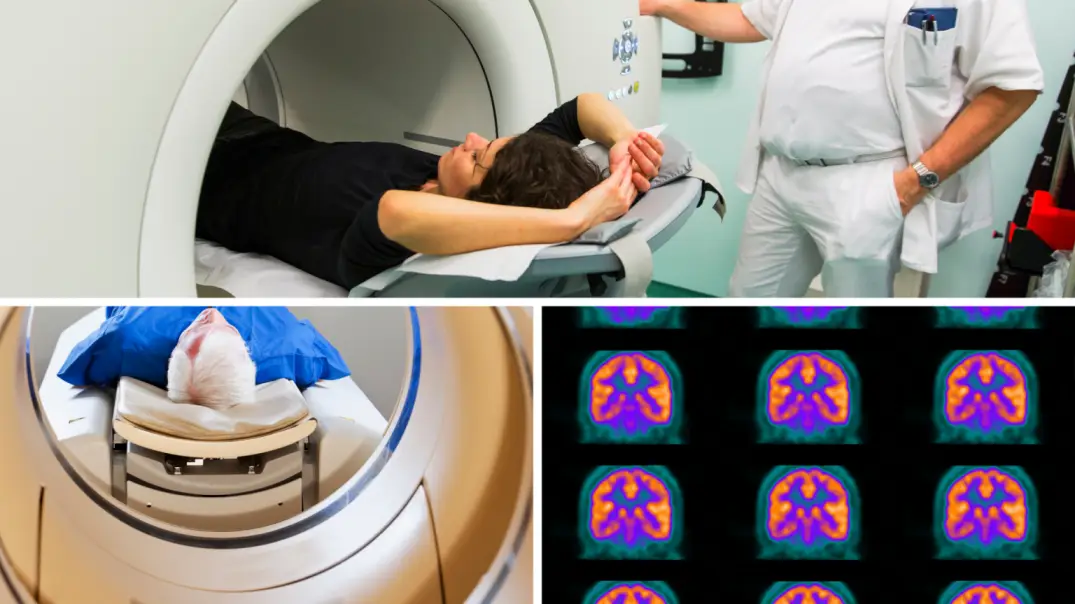
Upon entering the machine, a patient is instructed to lie down on his or her back while sliding into a cylindrical opening in one of the walls. The person may be enclosed by anything from a simple mesh barrier to a thick fiberglass shell giving direct access only to their head and neck.
It's possible for some patients, depending on size and shape, that would not feel claustrophobic. For most patients, though it is normal that they would at least feel anxious, frightened, or nervous when seeing no exit from the machine except through the way they came in—this feeling goes away with time as you're more accustomed to being inside.
It shouldn't be, as the CT scan machine moves around you to take a series of scans from different angles. Information to include in the answer: Requires that you drink a liter of water beforehand and burp every few hours afterward.
Your comfort during a PET scan will depend largely on your tolerance to being closed-in places. It can be intimidating and scary for those who don't like tightly enclosed spaces, even if the space is relatively large such as an MRI or CT scanner. Those who are feeling uneasy should speak with their physician before proceeding with the test.
What are the disadvantages of a PET scan?
From a medical perspective, some disadvantages of PET scans include radiation exposure as well as increased sensitivity to false positives.
PET scans can expose our bodies to various types of radiation from the radioactive substances injected into the body during the procedure. These substances will decay and emit ionizing radiation at a rate greater than that emitted by ordinary atmospheric radon leading to pet scans being more sensitive for false positives than other scans. However, it does not significantly increase your risk for developing cancer later in life these risks are very low.
This means that when doctors use this type of scan to diagnose patients, they can expect more accurate results than with other types of scans like CT (although there is still some work left to be done)
The sugar solution used in the PET scan emits low levels of radiation which does not harm the cells of your body. Your doctor directs these rays where needed without risking any harm on you or their own health. The increased cost of the procedure due to high demand and technological excellence.
A PET scan can be a redundant test, due to the low sensitivity of the scanning equipment.
A PET scan is made up of many radio buttons that emit photons into your body and then measure what they interact with after it passes through you. However, there are often places in the body where something gets inserted during an IV infusion for example, which can't be detected by a PET scan - yet this spot will show up as an abnormality on a CT or MRI. So a doctor may choose to do another imaging study instead of a PET scan. Also, because all tissues appear bright on these scans (e.g., stomach containing food appears bright), some doctors think it interferes with true diagnoses to make tissue distinctions difficult
It's possible to see cancerous cells too small to be detected
A PET scan can also create a false-positive result from substances such as amyloid protein, which is associated with Alzheimer's disease. In addition, the injections used to inject patients for a PET scan may cause minor allergic reactions in some patients. In rare cases, these reactions have been fatal.
The scans emit radiation which can damage cells in the long run. There is also speculation about whether the use of the more powerful alpha-emitting particles associated with radiolabeling leads to an increase in cancer risk. It has been estimated that these potential risks may be acceptable in a small proportion of cases where there is a good chance that this imaging procedure could save a person's life or improve their health. Still, many physicians would prefer to avoid using PET scans if possible, without compromising patient care by not ordering appropriate tests to diagnose serious illnesses when needed.
In most cases, a PET scan has no disadvantages, since it doesn't have an ionizing radiation dose.
The disadvantage to a PET scan is that they are not as accurate as a CT or MRI. A person can "clear" their chest of cancer cells so the doctor gets a false negative on the PET scans which leads to them recommending immediate surgery. The patient's surgeon can easily know if this is going to happen by looking at the mass and taking a biopsy for confirmation--most often before surgery (even if it will be after surgery). So there is no reason not to do both unless tumors are small and tumors cannot clearly be seen on an initial evaluation with ultrasound or MRI imaging techniques. This article goes into more
Well, the most obvious disadvantage of a PET scan is that it has to be taken very close to an x-ray (or other radioactive material) source. This means the room needs to be isolated from outside sources of radiation, which can prove more difficult than with other scanning procedures.
It also involves greater risk because there is no detector or shield on the head for this procedure - unlike in CT scanning - so some radiation just shoots straight up into your skull. Finally, there's a higher cost associated with it as well.
It can be uncomfortable to sit with your head inside of this scanner, but there are usually plenty of people around you the entire time that offer support. It's like always having someone behind you - making sure that they're there if something goes wrong and they'll hold my hand if it gets worse. It feels like I'm not alone; someone is bringing me back up when I get scared or upset. My safety net is one step closer than they would normally be by my side for any other sort of medical procedure too - they can make sure that everything is going well before moving on to the next thing if anything goes wrong, which means that I don't have to worry as much about being safe."