- Published on: Jul 30, 2024
- 4 minute read
- By: Secondmedic Expert
Ultrasound Imaging Explained: Benefits And Uses In Modern Medicine
Ultrasound imaging is a key tool in modern medicine, offering a safe and non-invasive way to view the inside of the body. By using high-frequency sound waves, ultrasound creates detailed images without radiation, making it invaluable for diagnosing and monitoring various conditions. In this blog, we’ll explore how ultrasound works, its benefits, and its common uses in healthcare. Join us as we uncover the role of ultrasound in enhancing patient care and advancing medical diagnostics.
What is Ultrasound Imaging?
Ultrasound imaging, also known as sonography, is a medical imaging technique that uses high-frequency sound waves to produce images of the inside of the body. Unlike X-rays or CT scans, ultrasound does not involve radiation, making it a safer option for various diagnostic procedures. This technology is widely used to examine organs, tissues, and blood flow, and it plays a crucial role in both routine and emergency medical assessments.
How Does Ultrasound Imaging Work?
Ultrasound imaging operates by emitting high-frequency sound waves through a transducer, a handheld device that looks like a small wand. Here’s a step-by-step look at the process:
-
Sound Wave Emission: The transducer sends high-frequency sound waves into the body. These sound waves travel through the body and bounce off different tissues and organs.
-
Echo Reception: The sound waves that reflect off tissues return to the transducer as echoes. The strength and timing of these echoes vary depending on the type of tissue they encounter.
-
Image Formation: The echoes are captured by the transducer and sent to a computer, which processes the data to create real-time images. These images display the internal structures of the body, allowing healthcare providers to view and assess them in detail.
-
Equipment Used: The primary equipment includes the transducer (or probe) and a computer with specialized software to generate and display the images. The transducer is often used with a gel applied to the skin to improve the transmission of sound waves.
By capturing and analyzing these sound wave reflections, ultrasound imaging provides valuable insights into the condition and function of internal organs, guiding diagnosis and treatment decisions.
Benefits of Ultrasound Imaging
-
Non-Invasive and Painless: Ultrasound imaging is a non-invasive procedure, meaning it does not require any incisions or needles. Patients typically experience no discomfort during the exam, making it a stress-free diagnostic tool.
-
No Radiation Exposure: Unlike X-rays or CT scans, ultrasound imaging uses sound waves instead of ionizing radiation. This makes it a safer option for frequent use, particularly in sensitive populations such as pregnant women and children.
-
Real-Time Imaging: Ultrasound provides real-time images, allowing healthcare providers to observe dynamic processes as they occur. This feature is crucial for assessing the function of organs and monitoring the progress of certain conditions.
-
Versatility: Ultrasound is a versatile imaging technique used for a wide range of diagnostic purposes. It can evaluate various body parts, from the abdomen to the heart, and is also effective in guiding certain medical procedures.
Common Uses of Ultrasound Imaging
-
Prenatal Care: Ultrasound is widely used in prenatal care to monitor the development of the fetus, check for any abnormalities, and determine the baby’s position and gender. It provides expectant parents with valuable information and reassurance throughout the pregnancy.
-
Cardiology: In cardiology, ultrasound is used to perform echocardiograms, which assess the heart's structure and function. This helps in diagnosing heart diseases, evaluating heart valve function, and guiding treatment decisions.
-
Abdominal and Pelvic Examinations: Ultrasound is used to examine organs in the abdomen and pelvis, such as the liver, kidneys, and bladder. It helps diagnose conditions like kidney stones, liver disease, and bladder abnormalities.
-
Musculoskeletal Imaging: This technique is effective in assessing muscles, tendons, and joints. It is often used to diagnose injuries, such as tears or sprains, and to guide treatment for musculoskeletal conditions.
-
Vascular Studies: Ultrasound is employed to study blood flow and detect issues in blood vessels, such as blockages or clots. This helps in managing conditions like deep vein thrombosis and assessing vascular health.
Ultrasound imaging’s broad applications and significant benefits make it an indispensable tool in modern medicine.
Advancements in Ultrasound Technology
Recent advancements in ultrasound technology have significantly enhanced its diagnostic capabilities and patient experience. Here are some key innovations:
-
3D and 4D Imaging: Modern ultrasound machines now offer 3D and 4D imaging capabilities, allowing for more detailed and dynamic views of internal structures. 3D imaging provides static, three-dimensional images, while 4D imaging adds the element of movement, which is particularly useful in prenatal care.
-
Doppler Ultrasound: This technology measures and visualizes blood flow within vessels and the heart. It helps in diagnosing conditions related to blood circulation, such as blockages or abnormalities in blood flow.
-
Portable Ultrasound Devices: Advances in technology have led to the development of portable ultrasound machines. These compact devices bring ultrasound capabilities to various settings, including remote or emergency locations, and enable quicker, on-the-go assessments.
-
Elastography: This technique assesses the stiffness of tissues, which can be crucial for diagnosing liver disease or tumors. Elastography provides additional information beyond traditional imaging, aiding in more accurate diagnoses.
-
Artificial Intelligence (AI): AI algorithms are increasingly integrated into ultrasound technology to enhance image quality, automate measurements, and assist in the interpretation of results. AI can improve diagnostic accuracy and streamline the imaging process.
These advancements make ultrasound imaging more versatile, accurate, and accessible, benefiting both patients and healthcare providers.
How to Prepare for an Ultrasound Examination
Preparation for an ultrasound examination can vary depending on the type of exam being performed. Here are some general tips to help ensure a smooth process:
-
Follow Specific Instructions: Your healthcare provider will give you specific instructions based on the type of ultrasound. For instance, you might need to fast for a period before an abdominal ultrasound or drink plenty of water before a pelvic exam.
-
Wear Comfortable Clothing: Choose clothing that allows easy access to the area being examined. For abdominal or pelvic ultrasounds, you may need to wear loose-fitting clothing.
-
Avoid Certain Foods or Drinks: If instructed to fast, avoid eating or drinking anything except water for the specified time. This helps ensure that your stomach and intestines are clear for the examination.
-
Arrive on Time: Arrive at your appointment a few minutes early to complete any necessary paperwork and to ensure that you’re prepared for the exam.
-
Inform the Technician: Let the ultrasound technician know if you have any medical conditions or if you are pregnant, as this may affect the imaging process.
Following these preparation tips will help ensure that your ultrasound examination is conducted smoothly and that the results are accurate.
Conclusion
Ultrasound imaging is a vital tool in modern healthcare, offering a non-invasive and radiation-free method for examining the body's internal structures. Its ability to provide real-time, detailed images makes it indispensable for diagnosing and monitoring a wide range of conditions, from prenatal assessments to cardiac evaluations. With recent advancements in technology, including 3D imaging and portable devices, ultrasound continues to enhance diagnostic accuracy and patient care.
By understanding and utilizing ultrasound imaging, healthcare providers can deliver more precise diagnoses and better manage patient treatment plans. This innovative technology not only supports effective medical decision-making but also contributes to improved patient outcomes and overall health management.
If you have any questions about ultrasound imaging or need to schedule an appointment, don’t hesitate to reach out to us at SecondMedic Healthcare. Our experienced team is here to provide you with the highest quality care and answer any inquiries you may have. Contact us today to learn more or to book your ultrasound examination!
Read FAQs
A. Ultrasound imaging, also known as sonography, uses high-frequency sound waves to create images of the inside of the body. A handheld device called a transducer emits these sound waves, which bounce off tissues and return as echoes. These echoes are then processed by a computer to produce real-time images of internal organs and structures.
A. Yes, ultrasound imaging is considered very safe. It does not use ionizing radiation, unlike X-rays or CT scans, making it a non-invasive and radiation-free diagnostic tool. It is commonly used in prenatal care to monitor the development of fetuses, demonstrating its safety and effectiveness.
A. During an ultrasound examination, a gel will be applied to your skin to help the transducer make secure contact and produce clear images. The technician will move the transducer over the area being examined. The process is typically painless and non-invasive. Depending on the type of exam, you might need to follow specific preparation instructions provided by your healthcare provider.
A. Ultrasound imaging is used for various diagnostic purposes, including prenatal care, cardiology, abdominal and pelvic examinations, musculoskeletal imaging, and vascular studies. It helps in monitoring fetal development, evaluating heart function, diagnosing abdominal and pelvic conditions, assessing musculoskeletal injuries, and examining blood flow in vessels.
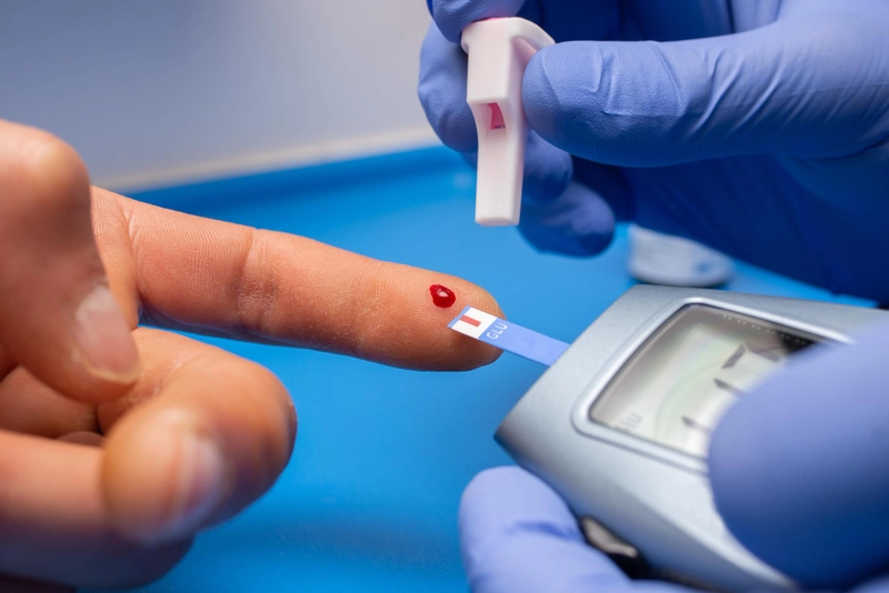
Reasons Behind Low Haemoglobin in Non-Anaemic People
When people think of low haemoglobin, they often assume it's always linked with anaemia. But that’s not always true. You can have a slightly low haemoglobin count even if your red blood cell count and iron levels are still in the normal range.
So, what does it mean when non-anaemic people have low haemoglobin? Should you worry? Let’s explore the reasons behind low haemoglobin in non-anaemic people and what you can do about it.
What Is Haemoglobin?
Haemoglobin is a protein found in red blood cells that carries oxygen from your lungs to the rest of your body. The average normal levels are:
-
Men: 13.5 – 17.5 g/dL
-
Women: 12.0 – 15.5 g/dL
Levels just below the normal range may not be classified as anaemia—but they can still indicate something going on.
Common Reasons for Low Haemoglobin in Non-Anaemic People
1. Mild Nutrient Deficiencies
Even if you’re eating regularly, you could still lack key nutrients needed to build haemoglobin—such as:
-
Vitamin B12
-
Folate (Vitamin B9)
-
Vitamin C (helps absorb iron)
Low levels of these don’t always lead to full-blown anaemia but can reduce haemoglobin production.
2. Chronic Inflammation or Infections
Your body may produce less haemoglobin during periods of chronic inflammation—such as:
-
Thyroid disorders
-
IBS or gut issues
-
Low-grade infections
These may not show symptoms right away but can slightly lower your haemoglobin over time.
3. Dilution from Overhydration
If you drink too much water before a blood test, your blood plasma volume may increase and dilute your haemoglobin, giving a lower reading.
This is temporary and often not harmful, but it can confuse test results.
4. Hormonal Imbalances
Conditions like hypothyroidism can subtly affect red blood cell and haemoglobin production.
In women, heavy menstrual bleeding can cause periodic dips in haemoglobin levels—especially if not supported with iron-rich nutrition.
5. Athletic Training (Pseudo-Anaemia)
In endurance athletes or those who do high levels of cardio, the body increases plasma (fluid) volume to improve circulation. This can lower the haemoglobin concentration without reducing red cell count—this is called athlete’s pseudo-anaemia.
What Tests to Consider
If you have low haemoglobin but no signs of anaemia, your doctor may recommend:
-
Serum Ferritin (iron storage)
-
Vitamin B12 and Folate tests
-
Thyroid profile
-
CRP or ESR (for inflammation)
You can get these tests easily with home sample collection from trusted platforms like SecondMedic.com, powered by Thyrocare.
Should You Be Concerned?
If your haemoglobin is:
-
Slightly below normal (e.g., 11.8–12.2 g/dL)
-
You have no symptoms (fatigue, paleness, breathlessness)
…then it may not be an emergency. However, monitoring and lifestyle changes are still important.
If it drops further or if symptoms appear, consult a doctor immediately.
What You Can Do Naturally
Improve Your Diet
-
Eat leafy greens, legumes, citrus fruits
-
Add iron-rich foods like beetroot, dates, and jaggery
-
Pair iron with vitamin C (e.g., lemon + spinach)
Reduce Inflammation
-
Avoid excessive sugar, fried foods, and processed meals
-
Include turmeric, ginger, and antioxidants in your diet
Stay Active but Balanced
-
Don’t overtrain
-
Rest and hydrate well, especially before blood tests
Conclusion
Low haemoglobin without anaemia is more common than you might think. The causes are often mild and reversible, but keeping an eye on your numbers and adjusting your diet or lifestyle can make a big difference.
If you’ve been wondering about the reasons behind low haemoglobin in non-anaemic people, now you know how to understand and manage it better—naturally and confidently.
Our Services
Request A Callback
Recent Posts
Lipid Profile Test – Normal Range and Risks
Jul 12,2025
How to Prevent Food Poisoning in Monsoon
Jul 10,2025
Aptamil Pepti Infant Formula 0 to 12 Months Tin 400gm
Brand: Nutricia International Private Limited
₹2000.00₹1960.00
View Details
Neocate LCP Infant Formula Powder 400GM
Brand: Nutricia International Private Limited
₹3300.00₹3234.00
View Details
Web Stories
Popular Lab Test
Live Doctor consultation
Live Doctor Chat
Fees ₹99
General Physician
Fees ₹99
General Physician
Fees ₹99
General Physician

Dr. Syed Mukhtar Mohiuddin
MBBS, MD , Fellowship in Diabetes Mellitus (UK)
- 7 Years
- English
Available at 00:00 to 07:00
Fees ₹99
General Physician
Fees ₹99
General Physician
Download Our App & Get Consultation from anywhere.