Prominent bronchovascular markings are a commonly reported finding on chest X rays and CT scans, yet they often cause confusion and concern when seen on a medical report. Many people come across this term unexpectedly and worry that it points to a serious lung condition.
In reality, bronchovascular markings are a normal part of lung anatomy. When they appear more visible than usual, it often reflects temporary changes such as inflammation, infection, or increased blood flow rather than permanent damage. The significance of prominent bronchovascular markings depends on symptoms, medical history, and whether other abnormalities are present on imaging.
Understanding what this finding means, what commonly causes it, and when it requires medical attention can help patients interpret their reports more confidently and avoid unnecessary anxiety.
What Are Bronchovascular Markings?
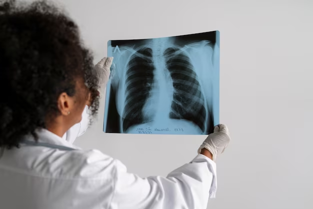
Bronchovascular markings are radiological features seen on chest imaging studies such as X rays and CT scans. They represent the branching patterns of the bronchi, which are the airways, and the blood vessels within the lungs.
These structures are a normal and necessary part of lung anatomy. Their role is to allow airflow and blood circulation throughout lung tissue so oxygen can be delivered efficiently to the body. Seeing bronchovascular markings on imaging is expected and does not by itself indicate disease.
How to Identify Bronchovascular Markings on Medical Images
On chest imaging, bronchovascular markings appear as fine lines or streaks extending outward from the central region of the lungs toward the periphery.
Bronchial markings usually appear slightly thicker and more defined, while vascular markings are finer and taper gradually as they move outward. This difference helps clinicians distinguish normal anatomical patterns from changes that may need further evaluation.
Visualizing Normal and Prominent Bronchovascular Markings
Understanding how bronchovascular markings look on imaging helps clarify why they are sometimes described as prominent.
Normal Bronchovascular Markings
-
The central lung region, known as the hilum, appears denser due to major airways and blood vessels
-
Fine branching lines extend outward and become thinner toward the lung edges
-
Lung margins and the diaphragm remain clearly defined
Prominent Bronchovascular Markings
-
Branching lines appear thicker or more noticeable than usual
-
Vascular markings may be more widespread and conspicuous
-
In some cases, lung margins or diaphragm outlines appear less distinct
Prominence is a descriptive observation and does not confirm a specific diagnosis on its own.
Reasons for Prominent Bronchovascular Markings
Prominent bronchovascular markings most commonly occur due to conditions that affect the airways, blood flow, or lung tissue. These causes range from temporary and mild to more persistent conditions.
Common Causes
Infections
Bacterial pneumonia and viral respiratory infections can cause inflammation and fluid accumulation in the lungs. This makes bronchovascular markings appear more visible on imaging.
Allergies and Asthma
Allergic reactions and asthma can trigger airway inflammation and increased mucus production, temporarily thickening bronchial walls.
Chronic Lung Conditions
Chronic obstructive pulmonary disease may cause long-term airway narrowing and mucus buildup. Bronchiectasis involves permanent airway dilation, often resulting in irregular or persistently prominent markings.
Other Potential Causes
Heart Conditions
Fluid buildup in the lungs related to heart failure can make vascular markings more pronounced.
Lung Tumors
In rare cases, tumors can alter normal airway or blood vessel patterns, usually alongside other imaging abnormalities.
Prominent Bronchovascular Markings Meaning in Clinical Context
When bronchovascular markings are described as prominent, it means they are more visible than expected on imaging. This description often reflects an underlying process such as inflammation or increased blood flow rather than a diagnosis.
In many individuals, especially those without ongoing symptoms, this finding is temporary and resolves once the underlying cause improves.
Is Prominent Bronchovascular Markings Serious?
The seriousness of prominent bronchovascular markings depends on the underlying cause and the presence of symptoms. In many cases, the finding is not serious.
Severity Levels and Associated Symptoms
Mild
Often related to minor infections or allergies, with symptoms such as mild cough or occasional wheezing.
Moderate
May be associated with chronic airway conditions, leading to persistent cough, mucus production, and shortness of breath during activity.
Severe
Can be linked to advanced lung or heart disease, with symptoms such as difficulty breathing at rest, chest tightness, or bluish skin discoloration.
The imaging term alone does not determine severity.
When to Seek Medical Advice
Medical attention is usually advised when prominent bronchovascular markings are accompanied by:
-
Persistent or worsening shortness of breath
-
Chest pain
-
Ongoing cough that does not improve
-
Fever lasting several days
-
Leg swelling or unexplained fatigue
These symptoms help healthcare providers determine whether further testing or follow-up imaging is needed.
What Usually Happens After This Finding Appears on a Report?
In many cases, no immediate treatment is required. Healthcare providers often correlate the imaging finding with symptoms and may recommend observation or repeat imaging, particularly if the scan was performed during an acute infection.
When symptoms persist or imaging changes progress over time, additional evaluation may be advised to identify and manage the underlying cause.
Treatment and Management
Treatment focuses on addressing the condition responsible for the imaging appearance rather than the bronchovascular markings themselves.
Lifestyle Changes
-
Smoking cessation to reduce airway irritation
-
Allergy management to minimize inflammation
-
Maintaining a healthy diet and regular physical activity
Medical Interventions
-
Medications to treat infections or reduce airway inflammation
-
Oxygen therapy in more severe respiratory conditions
-
Surgical intervention in rare cases, such as tumor removal
Preventing Prominent Bronchovascular Markings
While imaging findings cannot always be prevented, lung health can be supported through:
-
Regular medical checkups
-
Early treatment of respiratory infections
-
Avoiding smoking and environmental irritants
-
Proper management of chronic lung and heart conditions
Home Remedies for Mild Cases
For mild and temporary causes, supportive measures may help relieve symptoms:
-
Using a cool mist humidifier to loosen mucus
-
Drinking warm fluids to stay hydrated
-
Getting adequate rest and elevating the head during sleep
-
Avoiding allergens, smoke, and other irritants
These measures support recovery but do not replace professional medical care if symptoms worsen, consult a healthcare professional for a thorough evaluation and guidance.
Conclusion
Prominent bronchovascular markings on chest imaging are a descriptive finding, not a diagnosis. In many cases, they reflect temporary or manageable conditions rather than serious disease. Understanding their meaning, possible causes, and associated symptoms helps patients interpret imaging reports accurately and decide when medical evaluation is needed.